Holistic Balance

{Holistic Balance}{363 Burwood Rd}{Hawthorn}{3122}{VIC}{Australia}{(039) 041-3232}
363 Burwood Rd 3122 Hawthorn, VIC
Phone: (039) 041-3232
Blog | Holistic Balance - Part 3

Headaches

Author: Jerry Song

Headaches are one of the most common neurological complaints encountered in clinical practice, affecting individuals across various demographic groups. They can be broadly categorised into primary and secondary headaches, with each category encompassing multiple subtypes. Understanding the etiology of these headaches and the demographics most likely to suffer from them is crucial for effective diagnosis and management.


  1. Primary Headaches

Primary headaches are not caused by an underlying medical condition but are disorders themselves. The main types include migraines, tension-type headaches (TTH), and cluster headaches.

  1. Migraine

Migraine is a recurrent headache disorder characterised by moderate to severe pain, typically unilateral and pulsating in nature. The pain is often accompanied by nausea, vomiting, and sensitivity to light and sound. Migraines can be preceded by aura, which includes visual disturbances or other neurological symptoms.

Etiology: The exact cause of migraines is not fully understood, but it is believed to involve a combination of genetic, environmental, and neurological factors. There is evidence suggesting abnormal brain activity and blood flow changes, as well as dysfunction in the trigeminovascular system.

Demographics: Migraines affect approximately 12% of the global population, with a higher prevalence in women than men (about 3:1 ratio). They are most common in individuals aged 18 to 44, particularly among women of childbearing age due to hormonal fluctuations. There is also a genetic predisposition, as migraines tend to run in families.

  1. Tension-Type Headache (TTH)

Tension-type headaches are the most common type of primary headache. They are characterised by bilateral, mild to moderate pain, often described as a tight band around the head. These headaches can last from 30 minutes to several days.

Etiology: The etiology of TTH is believed to involve muscle tension and stress. Psychological factors such as anxiety and depression are also associated with TTH. The pain is thought to result from peripheral activation or sensitisation of myofascial nociceptors.

Demographics: Tension-type headaches affect up to 80% of the population at some point in their lives. They are slightly more common in women than men and are most frequently reported by individuals in their 30s and 40s. Stressful life situations and poor posture are common triggers.

  1. Cluster Headache

Cluster headaches are characterised by severe, unilateral pain, typically around the eye or temple, often accompanied by autonomic symptoms such as tearing, nasal congestion, or eyelid swelling. These headaches occur in clusters, with multiple attacks over weeks or months, followed by periods of remission.

Etiology: The exact cause of cluster headaches is unknown, but they are thought to involve abnormalities in the hypothalamus, leading to dysregulation of circadian rhythms. Genetic factors may also play a role, though they are less prominent than in migraines.

Demographics: Cluster headaches are rare, affecting about 0.1% of the population. They are more common in men than women (about 4:1 ratio) and typically begin between the ages of 20 and 40. Smoking and alcohol consumption are significant risk factors.

  1. Secondary Headaches

Secondary headaches are symptomatic of an underlying medical condition. The pain is often due to infection, trauma, vascular disorders, or other systemic issues. Common types include:

  1. Medication Overuse Headache (MOH)

Medication overuse headache, also known as rebound headache, occurs as a result of frequent use of headache medications. The headache is usually present upon waking and can resemble a tension-type headache or migraine.

Etiology: MOH is caused by the regular use of analgesics or triptans for headache relief, leading to a cycle of overuse and increased headache frequency.

Demographics: MOH is more prevalent in women, particularly those aged 30 to 50. It is also common among individuals with a history of chronic headaches, such as migraines or tension-type headaches.

  1. Sinus Headache

Sinus headaches are caused by inflammation or infection of the sinuses, leading to pressure and pain in the forehead, cheeks, and eyes. These headaches are often associated with sinusitis and may be accompanied by nasal congestion, fever, and facial swelling.

Etiology: Sinus headaches occur due to inflammation of the sinus cavities, often secondary to infection (viral or bacterial) or allergic reactions. The pain results from increased pressure within the sinuses.

Demographics: Sinus headaches are more common in individuals with a history of allergies or chronic sinusitis. They are equally prevalent among men and women and can affect all age groups, though they are more common in adults.

  1. Cervicogenic Headache

Cervicogenic headache is a secondary headache caused by disorders of the cervical spine and its components, such as intervertebral discs, facet joints, or cervical muscles. The pain is typically unilateral and may radiate from the neck to the front of the head.

Etiology: Cervicogenic headaches result from cervical spine disorders, such as osteoarthritis, herniated discs, or whiplash injuries. The pain is believed to originate from the convergence of nociceptive input from the cervical spine and trigeminal nerve.

Demographics: This type of headache is more common in individuals with neck trauma or chronic neck pain, particularly those aged 30 to 50. It is slightly more prevalent in women than men.


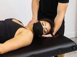
If you or someone you know are dealing with headaches, musculoskeletal pain, or even myofascial trigger points, book in an appointment to see Jerry now for an initial consultation so we can assess your condition and come up with an appropriate and tailored treatment plan to manage your pain. Simply click this booking link for the myotherapy initial consultation with our experienced myotherapists.

Compartment Syndrome

Compartment Syndrome 

Author: Jerry Song

Compartment syndrome is a condition where elevated pressure within a closed muscle compartment compromises circulation and function of the tissues within that space. It can be acute or chronic, with acute compartment syndrome (ACS) being a medical emergency requiring prompt surgical intervention. Chronic exertional compartment syndrome (CECS) is less urgent but can severely impact an individual’s ability to perform physical activities.

The causes of compartment syndrome vary based on whether it is acute or chronic:

  1. Acute Compartment Syndrome (ACS):
    • Trauma: Fractures, crush injuries, or severe contusions can lead to swelling or bleeding within a compartment.
    • Surgery: Postoperative complications can include bleeding or oedema causing elevated compartment pressures.
    • Vascular Injury: Trauma or surgery involving blood vessels can result in haemorrhage within a compartment.
    • Tight Casts or Dressings: External compression from casts or dressings can increase compartment pressures.
  2. Chronic Exertional Compartment Syndrome (CECS):
    • Repetitive Exercise: Activities involving repetitive muscle contractions, such as running or cycling, can increase compartment pressures over time.
    • Muscle Hypertrophy: Intense physical training leading to muscle hypertrophy can increase compartment size relative to its surrounding fascia, raising internal pressures.
    • Anatomic Abnormalities: Variations in fascial anatomy or muscle size can predispose individuals to CECS.


 Compartment syndrome affects various populations based on its type:

  1. Acute Compartment Syndrome:
    • Age: Can occur at any age but is more common in younger individuals due to higher incidence of trauma and sports injuries.
    • Gender: Males are more frequently affected, likely due to higher participation in high-risk activities and occupations.
    • Activities: Those involved in contact sports, high-impact activities, or occupations with a risk of severe injury are at increased risk.
  2. Chronic Exertional Compartment Syndrome:
    • Age: Commonly affects young athletes, typically in their late teens to early 30s.
    • Gender: Both genders are affected, but some studies suggest a higher incidence in females due to different biomechanics and muscle structures.
    • Sports and Activities: Athletes involved in repetitive, high-impact activities such as running, soccer, or military training are at greater risk.

Manual therapy plays a supportive role in managing chronic exertional compartment syndrome by alleviating symptoms, improving muscle function, and addressing contributing factors. Key manual therapy interventions include:

  1. Dry Needling

Dry needling involves the insertion of fine needles into specific trigger points or areas of muscle tightness to promote pain relief and muscle relaxation. Benefits of dry needling for compartment syndrome include:

  • Reduction of Muscle Tightness: Targeting trigger points in the affected muscles can alleviate tightness and improve muscle flexibility.
  • Pain Relief: By disrupting pain signals and promoting local blood flow, dry needling can effectively reduce muscle pain and discomfort.
  • Improved Muscle Function: Enhancing muscle function around the affected compartment can aid in reducing abnormal pressures and improving overall muscle performance.
  1. Myofascial Cupping

Myofascial cupping involves the use of suction cups to create negative pressure on the skin, lifting and separating the underlying tissues. This technique can:

  • Enhance Blood Circulation: Improved blood flow aids in nutrient delivery and waste removal, promoting tissue healing and reducing inflammation.
  • Release Myofascial Restrictions: Loosening tight fascia and muscle adhesions can restore normal tissue mobility and reduce pain.
  • Reduce Muscle Spasms: The negative pressure can relax muscle spasms, improving muscle tone and function within the affected compartment.
  1. Soft Tissue Mobilisation

Soft tissue mobilisation includes techniques such as massage and myofascial release to manipulate the soft tissues around the affected compartment:

  • Massage Therapy: Techniques like deep tissue massage can reduce muscle tension, improve circulation, and decrease pain, facilitating recovery of muscle function and flexibility.
  • Myofascial Release: Applying sustained pressure to myofascial restrictions can eliminate pain and restore motion, addressing tightness in the fascia and muscles.
  1. Joint Mobilisation

Joint mobilisation involves applying controlled forces to improve the range of motion and alleviate pain in the joints near the affected compartment:

  • Ankle and Knee Mobilisation: Techniques to mobilise the ankle and knee joints can enhance mobility, reduce stiffness, and correct abnormal mechanics contributing to compartment syndrome.
  1. Manual Stretching

Manual stretching techniques aim to improve flexibility and reduce tension in the muscles around the affected compartment:

  • Calf and Shin Stretching: Stretching the calf and shin muscles can relieve tension and improve muscle balance.
  • Quadriceps and Hamstring Stretching: Addressing tightness in the quadriceps and hamstrings can reduce stress on the affected compartment and improve overall limb function.
  1. Proprioceptive Neuromuscular Facilitation (PNF)

PNF stretching combines stretching and contracting targeted muscle groups to enhance flexibility and strength:

  • PNF Techniques: Effective for improving the flexibility and strength of the muscles within the affected compartment, promoting better mechanics and reducing pain.

If you or someone you know are dealing with Chronic Exertional Compartment Syndrome, musculoskeletal pain, or even myofascial trigger points, book in an appointment to see us now for an initial consultation so we can assess your condition and come up with an appropriate and tailored treatment plan to manage your pain. Simply click this booking link for the myotherapy initial consultation with our experienced myotherapists.


Femoroacetabular Impingement (FAI) Syndrome

Femoroacetabular Impingement (FAI) Syndrome

Femoroacetabular impingement (FAI) is a mechanical hip disorder resulting from abnormal morphological interactions between the femoral head and the acetabulum. This condition is often associated with hip pain, restricted movement, and the potential for early-onset osteoarthritis. FAI is classified into three types: cam, pincer, and mixed impingement, each defined by specific anatomical abnormalities.

FAI arises due to anatomical variations that cause abnormal contact and friction within the hip joint:

  1. Cam Impingement: Characterised by an aspherical femoral head that creates abnormal contact with the acetabulum during hip movements. This results in cartilage and labral damage.
  2. Pincer Impingement: Occurs when there is excessive coverage of the acetabulum over the femoral head, leading to labral compression and damage.
  3. Mixed Impingement: Combines features of both cam and pincer impingement, resulting in more complex pathomechanics and greater joint damage.

FAI commonly affects young and middle-aged adults, particularly those who are physically active. Specific demographics and risk factors include:

  1. Athletes: Individuals involved in sports requiring repetitive hip flexion, such as soccer, hockey, and dance, are at increased risk due to repetitive stress on the hip joint.
  2. Age and Gender: FAI typically presents in individuals aged 20-40 years. Males are more likely to develop cam impingement, while females are more prone to pincer impingement.
  3. Genetic Predisposition: Family history of hip disorders may increase the likelihood of developing FAI.
  4. Developmental Conditions: Conditions such as Legg-Calvé-Perthes disease or slipped capital femoral epiphysis during adolescence can predispose individuals to FAI.

Manual therapy is a critical component of FAI management, focusing on alleviating pain, improving hip mobility, and addressing muscular imbalances. Key manual therapy interventions include:

  1. Dry Needling

Dry needling involves the insertion of fine needles into specific trigger points or areas of muscle tightness to promote pain relief and muscle relaxation. Benefits of dry needling for FAI include:

  • Reduction of Muscle Tightness: Targeting trigger points in the hip flexors, adductors, and gluteal muscles can alleviate muscle tightness, enhancing hip mobility.
  • Pain Relief: By disrupting pain signals and promoting local blood flow, dry needling can effectively reduce hip pain.
  • Improved Muscle Function: Enhancing muscle function around the hip joint can aid in stabilising the joint and reducing abnormal stress.
  1. Myofascial Cupping

Myofascial cupping involves the use of suction cups to create negative pressure on the skin, lifting and separating the underlying tissues. This technique can:

  • Enhance Blood Circulation: Improved blood flow aids in nutrient delivery and waste removal, promoting tissue healing and reducing inflammation.
  • Release Myofascial Restrictions: Loosening tight fascia and muscle adhesions can restore normal tissue mobility and reduce hip pain.
  • Reduce Muscle Spasms: Cupping can relax muscle spasms, improving muscle tone and hip joint function.
  1. Soft Tissue Mobilisation

Soft tissue mobilisation includes techniques such as massage and myofascial release to manipulate the soft tissues around the hip:

  • Massage Therapy: Techniques like deep tissue massage can reduce muscle tension, improve circulation, and decrease pain, facilitating recovery of muscle function and flexibility.
  • Myofascial Release: Applying sustained pressure to myofascial restrictions can eliminate pain and restore motion, addressing tightness in the hip flexors, adductors, and iliotibial band.
  1. Joint Mobilisation

Joint mobilisation involves applying controlled forces to the hip joint to improve range of motion and alleviate pain:

  • Hip Joint Mobilisation: Techniques to mobilise the hip joint can enhance joint mobility, reduce stiffness, and correct abnormal joint mechanics.
  • Manual Traction: Applying gentle traction to the hip joint can reduce joint compression, alleviate pain, and improve joint space.
  1. Manual Stretching

Manual stretching techniques aim to improve flexibility and reduce tension in the muscles around the hip:

  • Hip Flexor Stretching: Stretching the hip flexors can relieve tension and improve hip extension.
  • Adductor Stretching: Addressing tightness in the adductors can restore balance and reduce stress on the hip joint.
  • Gluteal Stretching: Stretching the gluteal muscles can enhance hip mobility and stability.

Listed below are some details of a client dealing with FAIS who is seeking treatment here at HBM, which may also serve as a case study:

Age: 70+

Gender: F

Symptoms: Right Hip pain

Medical history: Arthritis

Lifestyle: Yoga at least once a week, walks and stands quite a lot while working.

The client has received 6 treatments here at HBM over the period of 6 months to manage her symptoms, and even after the first treatment has had great improvement in mobility and pain. Educating her on the importance of compliance to treatment due to her active schedule has made a big difference on her quality of life, and after educating her on the possible cause (after numerous questioning and orthopaedic testing) and with some take home exercises and stretches, she’s been able to get back to her daily activities with much improvements on symptoms.

If you or someone you know are dealing with FAIS, musculoskeletal pain, or even myofascial trigger points, book in an appointment to see us now for an initial consultation so we can assess your condition and come up with an appropriate and tailored treatment plan to manage your pain.

Simply click this booking link for the myotherapy initial consultation with our experienced myotherapists.


Repetitive strain injury (RSI)

Repetitive strain injury (RSI)

Repetitive strain injury (RSI) refers to a spectrum of conditions caused by repetitive movements and overuse of particular body parts, often leading to pain, discomfort, and functional impairment. RSIs are common in occupations and activities involving repetitive motions, prolonged exertion, or maintaining awkward positions.


RSI manifests in several forms, each affecting different anatomical structures:

  1. Carpal Tunnel Syndrome (CTS): Compression of the median nerve within the carpal tunnel of the wrist, leading to numbness tingling, and weakness in the hand.
  2. Tendinitis: Inflammation of tendons, commonly affecting the shoulders, elbows, and wrists. Examples include tennis elbow (lateral epicondylitis) and golfer’s elbow (medial epicondylitis)
  3. Bursitis: Inflammation of the bursae, small fluid-filled sacs that cushion bones and tendons, typically occurring in the shoulder, elbow and hip.
  4. De Quervain’s Tenosynovitis: Inflammation of the sheath surrounding the thumb tendons, causing pain and swelling at the base of the thumb.
  5. Thoracic Outlet Syndrome (TOS): Compression of nerves or blood vessels between the collarbone and first rib, causing pain in the neck, shoulders, and arms.


The primary causes of RSI include:

  • Repetitive Movements: Continuous, repetitive actions such as typing, using a mouse, or assembly line work.
  • Forceful Exertions: Activities requiring significant physical effort, including lifting heavy objects or using tools.
  • Sustained Postures: Maintaining fixed positions for extended periods, such as sitting at a desk or standing without adequate breaks.
  • Poor Ergonomics: Inadequate workstation setup, improper body mechanics, and poor posture.


Demographic Analysis:

RSI predominantly affects adults aged 25-50, particularly those in occupations involving repetitive tasks or prolonged static positions. Office workers, assembly line workers, musicians, and athletes are among the most vulnerable groups. The prevalence is higher in women, potentially due to differences in occupational roles and physical structure.

Manual Therapy encompasses a range of hands-on techniques performed by physical therapists or myotherapists to alleviate RSI symptoms. Key techniques include:

  • Massage therapy: Targets soft tissues to reduce muscle tension, improve circulation, and promote relaxation.
  • Joint mobilisations: Gentle, controlled movements of joints to restore range of motion and alleviate pain.
  • Soft tissue mobilisations: Techniques such as myofascial release to break down scar tissue and adhesions in muscles and connective tissues.
  • Trigger point therapy: Applying pressure to specific points to relieve muscle knots and referred pain.
  • Stretching and Strengthening exercises: Personalised exercises to improve flexibility, strength, and posture, reducing risk of future injury.

Listed below are some details of a client dealing with a shoulder RSI who is seeking treatment here at HBM, which may also serve as a case study:

Age: 35+

Gender: M

Symptoms: Shoulder pain after cricket match

Medical history: MRI showing slight oedema in subacromial bursa

Lifestyle: Sitting for work mainly, but workstation is not set up ergonomically; recently started playing cricket again.

The client has received 4 treatments here at HBM over 3 months to manage his symptoms, and even after the first treatment has had great improvement in mobility and pain. Educating him on the importance of compliance to treatment due to his active schedule has made a big difference on his quality of life, and after educating him on the possible cause (after numerous questioning and orthopaedic testing) and with some take home exercises and stretches, he’s been able to get back to his daily activities with much improvements on symptoms.

If you or someone you know are dealing with RSI, musculoskeletal pain, or even myofascial trigger points, book in an appointment to see us now for an initial consultation so we can assess your condition and come up with an appropriate and tailored treatment plan to manage your pain.

Simply click this booking link for the myotherapy initial consultation with our experienced myotherapists.


Temporomandibular Joint (TMJ) Syndrome

Temporomandibular Joint (TMJ) Syndrome

Temporomandibular Joint Syndrome (TMJ Syndrome), also known as temporomandibular disorder (TMD), is a condition affecting the temporomandibular joint (TMJ), which connects the jawbone to the skull. TMJ Syndrome is associated with pain, restricted jaw movement, and clicking or popping sounds in the jaw. The multifactorial nature of TMJ Syndrome makes its diagnosis and treatment challenging.

The development of TMJ Syndrome is attributed to various factors, including:

  1. Muscle Dysfunction: Overactivity or imbalance in the masticatory muscles can lead to pain and dysfunction in the TMJ.
  2. Joint Degeneration: Osteoarthritis or other degenerative joint diseases can affect the TMJ, leading to pain and limited movement.
  3. Trauma: Direct injury to the jaw or TMJ can result in acute or chronic TMJ Syndrome.
  4. Bruxism: Teeth grinding or clenching, often related to stress or anxiety, can cause excessive strain on the TMJ and surrounding muscles.
  5. Malocclusion: Misalignment of the teeth or bite can lead to abnormal stress on the TMJ, contributing to dysfunction.
  6. Psychosocial Factors: Stress, anxiety, and other psychological factors can exacerbate TMJ symptoms through increased muscle tension and altered pain perception.


TMJ Syndrome can affect individuals of all ages but is most commonly seen in adults aged 20-40 years. Specific demographics and risk factors include:

  1. Gender: Females are more likely to develop TMJ Syndrome than males, possibly due to hormonal influences and greater sensitivity to pain.
  2. Age: Young and middle-aged adults are more commonly affected, potentially due to higher stress levels and more frequent bruxism.
  3. Stress and Anxiety: Individuals experiencing high levels of stress or anxiety are at greater risk of developing TMJ Syndrome due to increased muscle tension and bruxism.
  4. Occupation and Lifestyle: Occupations and lifestyles involving high stress or repetitive jaw movements can predispose individuals to TMJ Syndrome.


Manual therapy is a critical component of TMJ Syndrome management, focusing on alleviating pain, improving jaw mobility, and addressing muscular imbalances. Key manual therapy interventions include:

  1. Dry Needling

Dry needling involves the insertion of fine needles into specific trigger points or areas of muscle tightness to promote pain relief and muscle relaxation. Benefits of dry needling for TMJ Syndrome include:

  • Reduction of Muscle Tightness: Targeting trigger points in the masticatory muscles, such as the masseter and temporalis, can alleviate muscle tightness and improve jaw function.
  • Pain Relief: By disrupting pain signals and promoting local blood flow, dry needling can effectively reduce jaw pain.
  • Improved Muscle Function: Enhancing muscle function around the TMJ can aid in stabilising the joint and reducing abnormal stress.
  1. Soft Tissue Mobilisation

Soft tissue mobilisation includes techniques such as massage and myofascial release to manipulate the soft tissues around the TMJ:

  • Massage Therapy: Techniques like deep tissue massage or trigger point therapy can reduce muscle tension, improve circulation, and decrease pain, facilitating recovery of muscle function and flexibility.
  • Myofascial Release: Applying sustained pressure to myofascial restrictions can eliminate pain and restore motion, addressing tightness in the jaw and neck muscles.
  1. Joint Mobilisation

Joint mobilisation involves applying controlled forces to the TMJ to improve range of motion and alleviate pain:

  • TMJ Mobilisation: Techniques to mobilise the TMJ can enhance joint mobility, reduce stiffness, and correct abnormal joint mechanics.
  • Manual Traction: Applying gentle traction to the TMJ can reduce joint compression, alleviate pain, and improve joint space.
  1. Manual Stretching

Manual stretching techniques aim to improve flexibility and reduce tension in the muscles around the TMJ:

  • Jaw Stretching: Stretching the jaw muscles can relieve tension and improve range of motion.
  • Neck and Shoulder Stretching: Addressing tightness in the neck and shoulder muscles can reduce referred pain and improve overall posture, benefiting TMJ function.
  1. Proprioceptive Neuromuscular Facilitation (PNF)

PNF stretching combines stretching and contracting targeted muscle groups to enhance flexibility and strength:

  • PNF Techniques: Effective for improving the flexibility and strength of the jaw and neck muscles, promoting better joint mechanics and reducing pain.

Listed below are some details of a client dealing with TMJ syndrome who is seeking treatment here at HBM, which may also serve as a case study:


Age: 50+

Gender: F

Symptoms: Left TMJ pain

Medical history: None pertinent

Lifestyle: Seated for work mostly. Does Kieser 3x/week as exercise and does a bit of jogging.

The client has been receiving fortnightly treatments here at HBM over a period of 3 months to manage her symptoms, and even after the first treatment has had great improvement in mobility and pain. Educating her on the importance of compliance to treatment due to her active schedule has made a big difference on her quality of life, and after educating her on the possible cause (after numerous questioning and orthopaedic testing) and with some take home exercises and stretches, she’s been able to get back to her daily activities with much improvements on symptoms.

If you or someone you know are dealing with TMJ syndrome, musculoskeletal pain, or even myofascial trigger points, book in an appointment to see us now for an initial consultation so we can assess your condition and come up with an appropriate and tailored treatment plan to manage your pain.  Simply click this booking link for the myotherapy initial consultation with our experienced myotherapists.


Tennis elbow Vs Golfer’s elbow

Tennis elbow and golfer’s elbow are overuse injuries that result from repetitive stress on the tendons and muscles around the elbow joint, leading to microtrauma, inflammation, and tissue degeneration. Despite their distinct anatomical locations, both conditions share common risk factors and clinical manifestations, including pain, tenderness, and functional impairment.

Tennis elbow, or lateral epicondylitis, involves inflammation and degeneration of the tendons attached to the lateral epicondyle of the humerus, primarily affecting the extensor muscles of the forearm responsible for wrist extension and radial deviation. In contrast, golfer’s elbow, or medial epicondylitis, affects the tendons attached to the medial epicondyle of the humerus, involving the flexor muscles of the forearm responsible for wrist flexion and ulnar deviation. While both conditions result from repetitive stress, they differ in their biomechanical mechanisms, anatomical involvement, and specific activities that exacerbate symptoms.

Photo illustration


Individuals engaged in activities requiring repetitive wrist and forearm movements are at increased risk of developing tennis and golfer’s elbow. Specific demographic groups susceptible to these conditions include:

  • Athletes: Tennis players, golfers, baseball players, and other athletes engaging in racquet sports or throwing activities are prone to developing tennis or golfer’s elbow due to the repetitive nature of their sport and high-intensity wrist and forearm movements.
  • Manual Laborers: Workers involved in occupations such as construction, plumbing, carpentry, and painting are at risk of developing tennis or golfer’s elbow due to the repetitive use of hand tools and sustained wrist positions.
  • Office Workers: Individuals performing repetitive typing, mouse clicking, and other computer-related tasks are susceptible to tennis or golfer’s elbow, as prolonged wrist extension or flexion can exacerbate symptoms.
  • Age and Gender: Middle-aged individuals are more commonly affected by tennis and golfer’s elbow, although these conditions can occur in individuals of all ages. Golfer’s elbow tends to be more prevalent in men, while tennis elbow affects both men and women equally.

Elbow Pain

Elbow Pain 2


Manual therapy interventions play a crucial role in the conservative management of tennis and golfer’s elbow, aiming to reduce pain, improve tissue healing, and restore functional mobility. Common manual therapy techniques employed in the treatment of these conditions include:
• Soft Tissue Mobilisation: Manual manipulation of the muscles, tendons, and fascia surrounding the affected elbow aims to reduce adhesions, improve tissue extensibility, and alleviate pain and inflammation.
• Joint Mobilisation: Gentle mobilisation of the elbow joint and adjacent joints, including the wrist and shoulder, can help restore normal joint mechanics, reduce stiffness, and optimise biomechanical function.
• Stretching and Strengthening Exercises: Specific exercises targeting the muscles of the forearm, wrist, and shoulder aim to improve flexibility, strength, and neuromuscular control, thereby reducing strain on the affected tendons and promoting tissue healing.

Listed below are some details of a client dealing with tennis elbow who is seeking treatment here at HBM, which may also serve as a case study:

Age: 35+
Gender: M
Symptoms: pain in R elbow from tennis 3x/week
Medical history: MRI showing disc herniation in C6-7
Lifestyle: Plays tennis 3x/week 2 hrs each for the past 2 years. Doesn’t stretch much and isn’t very flexible.

The client has received fortnightly treatments here at HBM over 8 months to manage his symptoms, and even after the first treatment has had great improvement in mobility and pain. Educating him on the importance of compliance to treatment due to his active schedule has made a big difference on his quality of life, and after educating him on the possible cause (after numerous questioning and orthopaedic testing) and with some take home exercises and stretches, he’s been able to get back to his daily activities with much improvements on symptoms.

If you or someone you know are dealing with tennis or golfer’s elbow, musculoskeletal pain, or even myofascial trigger points, book in an appointment to see us now for an initial consultation so we can assess your condition and come up with an appropriate and tailored treatment plan to manage your pain.

Simply click this booking link for the myotherapy initial consultation with our experienced myotherapists.

Carpal Tunnel Syndrome

Carpal tunnel syndrome (CTS) is a common peripheral nerve disorder resulting from compression of the median nerve as it passes through the carpal tunnel, a narrow passageway in the wrist formed by the carpal bones and transverse carpal ligament. The compression of the median nerve leads to a constellation of symptoms, including pain, numbness, tingling, and weakness in the thumb, index, middle, and radial half of the ring finger. While CTS can have multifactorial etiology, manual therapy has emerged as a promising adjunctive treatment modality to alleviate symptoms and improve hand function.

Hand illustration The pathophysiology of CTS is complex and involves mechanical compression, ischemia, and inflammation of the median nerve within the carpal tunnel. Predisposing factors such as wrist anatomy, repetitive hand movements, trauma, hormonal changes, and systemic diseases contribute to the development of CTS. Compression of the median nerve leads to impaired nerve conduction, demyelination, and axonal degeneration, resulting in sensory and motor deficits in the affected hand.

CTS predominantly affects individuals engaged in repetitive hand and wrist movements, as well as those exposed to occupational or recreational activities that involve prolonged wrist flexion or extension. Thus, certain demographic groups are more susceptible to developing CTS, including:• Working Professionals: Occupations that involve repetitive or forceful hand movements, prolonged computer use, or vibrating tools predispose individuals to CTS. Office workers, assembly line workers, musicians, and those in the construction industry are at increased risk.
• Women: Studies have shown that women are more likely to develop CTS compared to men, possibly due to hormonal factors, smaller carpal tunnel dimensions, and increased susceptibility to soft tissue swelling during pregnancy.
• Aging Population: Older adults are at higher risk of developing CTS due to age-related changes in soft tissues, decreased nerve elasticity, and underlying degenerative conditions such as arthritis and ligamentous laxity.
• Individuals with Comorbidities: Systemic conditions such as diabetes, obesity, thyroid disorders, and rheumatoid arthritis are associated with an increased risk of developing CTS due to their effects on nerve health, tissue inflammation, and fluid retention.

Manual therapy encompasses a variety of hands-on techniques aimed at addressing musculoskeletal dysfunctions, improving joint mobility, and reducing soft tissue restrictions. In the context of CTS, manual therapy interventions target the underlying anatomical and biomechanical factors contributing to median nerve compression. Common manual techniques employed in the treatment of CTS include:
• Soft Tissue Mobilisation: Manual manipulation of the muscles, tendons, and fascia surrounding the carpal tunnel aims to reduce adhesions, improve tissue extensibility, and alleviate pressure on the median nerve.
• Nerve Gliding Exercises: Specific exercises designed to mobilise and stretch the median nerve can help reduce neural tension, improve nerve conduction, and alleviate symptoms of CTS.
• Joint Mobilisation: Gentle mobilisation of the wrist, hand, and adjacent joints aims to improve joint mobility, reduce stiffness, and optimise the biomechanics of the upper extremity.
• Therapeutic Exercises: Strengthening and stretching exercises targeting the muscles of the forearm, hand, and wrist can help improve grip strength, enhance proprioception, and promote functional recovery in individuals with CTS.

Hand Illustration If you or someone you know are dealing with CTS, musculoskeletal pain, or even myofascial trigger points, book in an appointment to see us now for an initial consultation so we can assess your condition and come up with an appropriate and tailored treatment plan to manage your pain.

Simply click this booking link for the myotherapy initial consultation with our experienced myotherapists.

Thoracic Outlet Syndrome

Thoracic outlet syndrome (TOS) encompasses a group of disorders caused by compression of the neurovascular structures, including the brachial plexus, subclavian artery, and vein, as they pass through the thoracic outlet. The thoracic outlet is anatomically defined as the passageway between the clavicle and first rib, bounded by the scalene muscles anteriorly, the first rib inferiorly, and the posterior cervical muscles posteriorly. TOS can be classified into neurogenic, vascular, or nonspecific types, each presenting with distinct clinical features.

TOS can arise from various anatomical anomalies or acquired conditions that result in compression of the thoracic outlet structures. Common etiological factors include congenital anomalies such as cervical ribs, abnormal scalene muscle anatomy, muscular hypertrophy, trauma, poor posture, repetitive overhead activities, and anatomical variations in the bony and soft tissue structures. The compression leads to ischemia, nerve irritation, and mechanical distortion, giving rise to the characteristic symptoms of TOS.

Patients with TOS typically present with a constellation of symptoms, including pain, numbness, tingling, and weakness in the upper extremities. Neurogenic TOS primarily manifests with neurological symptoms, such as paresthesia along the ulnar aspect of the forearm and hand, while vascular TOS presents with symptoms related to arterial or venous compromise, such as cold intolerance, pallor, and swelling. Nonspecific TOS may exhibit a combination of neurogenic and vascular symptoms, making diagnosis challenging and often necessitating a multidisciplinary approach.

Manual therapy encompasses a variety of hands-on techniques aimed at restoring mobility, reducing pain, and improving function in musculoskeletal disorders. In the context of TOS, manual therapy interventions target the underlying musculoskeletal and soft tissue dysfunctions contributing to thoracic outlet compression. Techniques commonly employed include:

  • Soft Tissue Mobilisation: Manual manipulation of soft tissues, such as massage, myofascial release, and trigger point therapy, aims to alleviate muscular tension, reduce adhesions, and improve blood flow to the affected area.
  • Joint Mobilisation: Gentle mobilisation of the thoracic spine, cervical spine, and ribs can help restore normal joint mechanics, alleviate restrictions, and reduce nerve impingement.
  • Stretching and Strengthening Exercises: Specific exercises targeting the muscles of the neck, shoulder girdle, and upper extremities can improve flexibility, strength, and postural alignment, thereby reducing compression on the thoracic outlet structures.
  • Postural Correction: Education and training in proper posture and ergonomics are essential components of manual therapy for TOS, as poor posture contributes to thoracic outlet compression and exacerbates symptoms.

Listed below are some details of a client dealing with pain associated with TOS who is seeking treatment here at HBM, which may also serve as a case study:

Age: 40+

Gender: M

Symptoms: Nerve pain in elbow down to index finger on R hand

Medical history: No known injuries of relevance.

Lifestyle: Travels abroad frequently for work, while finishing a PhD at the same time.

The client has received 5x treatments here at HBM over 5 months to manage his symptoms, and even after the first treatment has had great improvement in mobility and pain. Educating him on the importance of compliance to treatment even with such a busy schedule has made a big difference on his quality of life, and after educating him on the possible cause (after numerous questioning and orthopaedic testing) and with some take home exercises and stretches, he’s been able to get back to his daily activities with much improvements on symptoms.

If you or someone you know are dealing with TOS, musculoskeletal pain, or even myofascial trigger points, book in an appointment to see us now for an initial consultation so we can assess your condition and come up with an appropriate and tailored treatment plan to manage your pain.

Simply click this booking link for the myotherapy initial consultation with our experienced myotherapists.

Lower Cross Syndrome

Lower Cross Syndrome


Lower crossed syndrome (LCS) is a musculoskeletal imbalance condition characterised by specific patterns of muscle dysfunction, primarily involving the lumbar spine, pelvis, and lower extremities. Tightness in the hip flexors (iliopsoas, rectus femoris) and lumbar extensors (erector spinae) is coupled with weakness in the gluteal muscles and abdominal stabilisers (transversus abdominis, internal obliques). The imbalanced muscle activity results in an anterior pelvic tilt, increased lumbar lordosis, and a protruding abdomen, leading to a characteristic swayback posture.

Individuals with LCS often experience chronic low back pain, hip pain, and tightness in the hip flexors. Dysfunction in the lumbar-pelvic region may contribute to secondary symptoms such as sacroiliac joint dysfunction and hip joint impingement. Additionally, LCS can impair functional movement patterns, affecting activities such as walking, standing, and squatting. Reduced hip mobility and stability may also predispose individuals to compensatory movements and increased risk of injury during physical activities.

One method myotherapists may use to address LCS is by manual therapy such as soft tissue mobilisation techniques, which include myofascial release and trigger point therapy, that target tight muscles (e.g., hip flexors, lumbar extensors) to alleviate tension and improve flexibility. Joint mobilisation and manipulation may also be employed to restore normal joint mechanics and address segmental dysfunctions, particularly in the lumbar spine and pelvis.


In addition to manual therapy techniques, exercise therapy is very beneficial and complements the above techniques in addressing LCS more comprehensively. Strengthening exercises focusing on the gluteal muscles and abdominal stabilisers are essential components of LCS rehabilitation. Hip extension exercises, core stabilisation exercises, and postural retraining aim to correct muscle imbalances and promote optimal alignment of the lumbar-pelvic region.

Listed below are some details of a client dealing with pain associated with LCS who is seeking treatment here at HBM, which may also serve as a case study:

Age: 40+

Gender: F

Symptoms: Immense pain around lower back suddenly after going down the stairs one day, could barely walk after it happened.

Medical history: No significant injuries shown on MRI/X-Ray

Lifestyle: Works as a music teacher, and is seated for that mostly, also does Pilates to strengthen her muscles

The client has received 3x treatments here at HBM over 1.5 months to manage her symptoms, and even after the first treatment has had great improvement in mobilityand pain. Educating her on the importance of compliance to treatment has made a big difference on her quality of life, and after educating her on the possible cause (after numerous questioning and orthopaedic testing) and with some take home exercises and stretches, she’s been able to get back to her daily activities relatively pain-free.

If you or someone you know are dealing with LCS, musculoskeletal pain, or even myofascial trigger points, book in an appointment to see us now for an initial consultation so we can assess your condition and come up with an appropriate and tailored treatment plan to manage your pain.

Simply click this booking link for the myotherapy initial consultation with our experienced myotherapists.

The Overlooked Facts of Fascia

Fascia is a complex, three-dimensional network of connective tissue that pervades the entire human body, surrounding and interpenetrating muscles, bones, nerves, and organs. Fascia consists of collagen fibers, elastin, ground substance, and cells such as fibroblasts. The arrangement of these components contributes to the diverse mechanical properties of fascial tissues. Fascia serves as a dynamic system with several key functions, including providing structural support, facilitating movement and flexibility, and participating in proprioception. Moreover, it acts as a conduit for neurovascular structures and plays a vital role in intercellular communication.

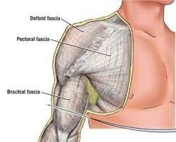
Some of the common fascial conditions include Myofascial Pain Syndrome (MPS), which is a pathology characterised by the development of trigger points within fascial tissues. These trigger points can lead to localised or referred pain, muscle stiffness, and reduced range of motion. Another fascial condition that may arise is fascial adhesions, which occur when collagen fibers in the fascial matrix become distorted or adhered, resulting in restricted movement and impaired tissue glide. This can contribute to chronic pain and dysfunction.


The Role of Manual Therapy in Fascial Treatment:


  1. Soft Tissue Mobilisation:

Manual therapy techniques, such as soft tissue mobilisation, aim to address fascial restrictions by applying controlled pressure and movement to specific areas. This helps to break down adhesions, improve tissue flexibility, and restore normal function.

  1. Myofascial Release:

Myofascial release involves sustained pressure and stretching of fascial tissues to release tension and promote optimal tissue mobility. Therapists use various hands-on techniques to target specific areas of fascial restriction.

  1. Instrument-Assisted Soft Tissue Mobilisation (IASTM):

IASTM employs specialised tools to detect and treat fascial dysfunction. By applying controlled pressure through these instruments, therapists can break down adhesions and enhance the healing response.

Aside from the above, myofascial cupping and dry needling are two therapeutic modalities that can be effective in addressing fascial conditions, contributing to improved tissue mobility, pain relief, and overall well-being.

Cupping involves placing suction cups on the skin, creating a negative pressure environment. This negative pressure lifts the fascial layers, increasing blood flow and promoting the release of fascial restrictions. The suction effect can also create a separation between the layers of fascia, reducing adhesions and facilitating better tissue glide. Cupping is believed to also stimulate the lymphatic system, aiding in the removal of toxins and metabolic waste from the fascial tissues.

Dry needling involves the insertion of thin, solid needles into trigger points or tight bands of fascial tissue. These trigger points are areas of hyperirritability within the fascia. The needling creates a microtrauma, prompting a local twitch response in the muscle fibers. This response helps release tension and reset the neuromuscular system.

Listed below are some details of a client dealing with lower back pain who is seeking treatment here at HBM, which may also serve as a case study:

Age: 40+

Gender: M

Symptoms: Sensation of tightness in the lower back and forearms

Medical history: Disc bulge injury in L4-S1 in the past, Tennis elbow

Lifestyle: Works at a desk and sits in front of a screen for long periods. Did a lot of boxing until recently, when he increased his weightlifting sessions.

The client has received 3x treatments here at HBM over 1.5 months to manage his symptoms, and even after the first treatment has had great improvement in mobility. Educating him on the importance of compliance to treatment has made a big difference on his quality of life.

If you or someone you know are dealing with lower back pain, tennis elbow, headaches/migraines, and think it may be due to a fascial condition, book in an appointment to see us now for an initial consultation so we can assess your condition and come up with an appropriate and tailored treatment plan to manage your pain.

Simply click this booking link for the myotherapy initial consultation with our experienced therapists.


Review

Google Rating
4.9
Based on 305 reviews
js_loader